Table of Contents
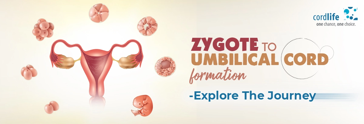
“The wonder of life begins in a woman’s womb.” — Lailah Gifty Akita. This is a beautiful feeling, right? But, do you know the core behind this beauty? Well! Everything begins with the fertilisation of an egg.
After the egg fertilises, the development of blastocyst, embryo, and fetus takes place.
In this article, we’re going to focus on each development elaborately and carefully.
Egg Fertilisation and Zygote Formation
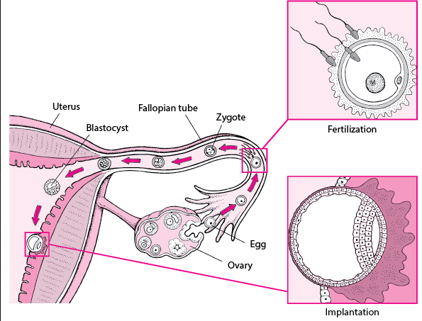
During each normal menstrual cycle, an egg (ovum) is released from each of the ovaries approximately 14 days after the last menstrual period. This process is known as ovulation. Subsequently, the egg travels into the funnel-shaped end of one of the fallopian tubes.
For the fertilisation of the eggs, the sperm have a big role to play. So, during ovulation, the consistency of the mucus in the cervix changes. It becomes more fluid and more elastic. Due to this change, after sexual intercourse, the sperm enters the uterus easily. How?
Remarkably, within 5 minutes, the sperm travels from the vagina, through the cervix and then reaches the uterus. Eventually, it reaches the funnel-shaped end of one of the fallopian tubes to facilitate the process of fertilisation. That said, egg fertilisation occurs when the sperm penetrates the egg successfully gradually making its way to the uterus. The fertilisation process completes in 24 hours and the fertilised egg is known as a zygote.
Zygote Formation and Blastocyst Development
The fertilised ovum or egg also known as the zygote, is formed because of the fusion of two cells – sperm and an egg. Initially, the zygote exists as a single cell, and remarkably, this single cell has a complete set of 46 chromosomes. Sperms contribute 23 cells and eggs contribute 23. But, after egg fertilisation, this single cell goes through several divisions and enables the zygote to multiply and develop further. Having said that, the zygote phase lasts only for four days. (It travels down the fallopian tube and enters the uterus in 3 to 5 days). Around the 5th day, the cluster of cells or solid ball of cells transforms or develops into a blastocyst. By nearly the 9th or 10th day, the blastocyst attached itself to the inner wall of the uterus, further allowing it to develop into an embryo.
The outer cells, however, develop into the placenta and have a huge role to play in maintaining pregnancy and developing the foetus, besides carrying the oxygen and nutrients from the mother to the foetus and removing waste from the foetus to the mother.
That said, specific cells from the placenta go through specialisation and can give rise to the outer layer of membranes, which is called the chorion. At the same time, the other cells develop into an inner layer of membranes called amnion which forms the amniotic sac. Gradually, the amniotic sac, filled with a clear fluid expands to enclose and support the developing embryo and the embryo floats within a safe environment.
Embryonic and Foetal Development
The embryo starts to grow within the amniotic sac nestled beneath the uterine wall. At this stage, most of the internal as well as external organs start forming. Around three weeks after egg fertilisation, the embryo starts to elongate and take a rudimentary human shape. Shortly after that, the brain and the spinal cord develop. Within the 20th day, the major blood vessels and heart begin developing. The heart also starts circulating fluid through blood vessels and the first red blood cell emerges the following day.
By around 10 weeks after fertilisation, all the organs may be formed, but the brain, as well as the spinal cord, continue to grow throughout the pregnancy journey.
By the 8th week, however, the embryo with almost all the organs becomes a foetus. In fact, the foetus will cover the entire uterus, by 12 weeks. The foetal sex will get identified, by around 14 weeks. From 16 to 20 weeks the foetus will start moving. Around the 24-week mark, the fetus reaches a stage where there is a possibility of survival outside the uterus. The foetal lungs continue to develop till such time that you give birth to the baby. In fact, the brain develops new cells throughout the pregnancy journey.
As the placenta develops, at this stage, it generates small hairlike projections called villi, which extend into the uterine wall. Within these villi, blood vessels originating from the embryo and passing through the umbilical cord to the placenta begin to form.
Umbilical Cord Development
The umbilical cord is the most essential connection between the placenta and the baby developing inside you. The formation of the connecting stalk around the 3 weeks, develops the umbilical cord, however, by the 7th week, the cord that connects you to your baby, is fully formed. The umbilical cord is composed of the umbilical vessels, connecting stalk, and vitelline duct. The umbilical vessels and arteries have a crucial role to play in the exchanging of oxygen, nutrients, and waste products during pregnancy. The average length of an umbilical cord is 50 to 60 cm.
Once you deliver your baby, the umbilical cord is cut and clamped to separate you from your baby. And if you’ve decided on cord blood banking because cord blood is rich with blood-forming stem cells, get ready.