Table of Contents
- Conception and Formation of the Zygote and Umbilical Cord
- Step 1: Conception (Day 0)
- Step 2: The Cellular Road Trip (Days 1-4)
- Step 3: The Blastocyst: Finding a Home and Settling In
- Step 4: Gastrulation: Building the Body’s Three Master Layers
- Step 5: Morphogenesis: Shaping the Body and Forming the Neural Tube:
- Step 6: Formation of the Placenta and Umbilical Cord
- Step 7: The Foetal Transition – From Building to Growing
- Conclusion
The miraculous journey of a single microscopic cell into a completely new human being is truly the most incredible of origin stories. This journey shows us exactly how life develops, from the first cellular division to the formation of the umbilical cord and placenta. Long before a mother can feel that first foetal movement, or monitor her baby’s heartbeat, that cell starts its journey from zygote to umbilical cord development. It is from this cellular beginning that a tiny baby takes shape. This article charts how one cell takes its first amazing steps towards creating a baby!
Conception and Formation of the Zygote and Umbilical Cord
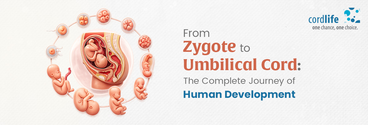
The journey of human development from zygote to foetus starts with the most exclusive cellular adventure tour. Once the remarkable moment of conception has occurred, a single cell begins navigating the fallopian tube, preparing for one of the most extraordinary feats in the body, dividing repeatedly without increasing significantly in overall size.
The following steps explain how this microscopic voyage unfolds:
Step 1: Conception (Day 0)
Somewhere within the upper, outermost part of the fallopian tubes, one tiny sperm penetrates the outer layer surrounding the egg. The egg then undergoes changes that prevent other sperm from entering. The genetic matter from the dad’s sperm and the mom’s egg merge to recreate the standard set of 46 chromosomes that will guide the development of the baby.
The Zygote is born
The brand-new, single-celled zygote comes into existence! This perfect, new single cell already carries everything. The Zygote has the ultimate plan, which encompasses each detail from eye and hair color approximate size of the body, to the type of digestive enzymes it will produce.
Step 2: The Cellular Road Trip (Days 1-4)
The zygote journeys down to the comfort and warmth of the mother’s uterus. On its way, it begins the construction of the future baby.
So how does a zygote develop into a baby?
It undergoes a very special type of cell division called cleavage over the next few days. The single original cell splits into many cells, but the developing structure remains relatively small in size.
Imagine a piece of clay being pinched in two, then four pieces, then eight.
Day 1 (24 to 36 hours):
The Zygote finishes the first round of division, producing a tidy 2-cell package still tucked into its protective envelope.
Day 2 or 3 (48 to 72 hours):
Many clone cells continue to divide, and gradually 4-cell or 8-cell structures develop into a 16-cell cluster. At this stage of early pregnancy development, each cell has the capability of forming into any type of human tissue or the placenta!
Day 4 (96 hours):
By the fourth day, the Zygote develops into a tightly packed ball of about 16-32 cells, commonly referred to as a ‘morula’, resembling a mulberry. This tiny cluster is swept down the fallopian tube and is propelled towards the uterus, ready to nest.
Step 3: The Blastocyst: Finding a Home and Settling In
On day 5, the travelling cluster reaches the warm, cosy uterus. As the cells arrive, fluid begins to pool right in the centre of the cell mass, expanding it into a hollow sphere called a blastocyst. The blastocyst has two main parts:
- The inner cell mass: This will become the baby
- The outer layer of cells: Known as the trophoblast, these cells contribute to the formation of the placenta.
Finally, on day 6 or 7, the blastocyst nestles in the warm uterine wall. This process of implantation creates the initial blood link between the embryo and its mother, and jump-starts the embryo development timeline. [8] [9]
Step 4: Gastrulation: Building the Body’s Three Master Layers
From the third and fourth week onwards, the cells remodel into 3 layers known as primary germ layers.
- The outermost layer, the ectoderm: This later becomes the skin, hair, nails, and entire nervous system, including the brain and spinal cord.
- The middle layer, the mesoderm: This forms the structural components, namely the skeleton, skeletal muscles, heart and vascular system, kidneys, etc.
- The innermost layer, the endoderm: This develops into the digestive system, liver, pancreas, lungs, and respiratory system.
Gastrulation is a crucial stage of human development during pregnancy, when the basic framework for the body’s major systems begins to take shape.
Step 5: Morphogenesis: Shaping the Body and Forming the Neural Tube:
The embryo embarks on an amazing journey from weeks 3 to 8. The organs take shape, and the nervous system starts forming. The uppermost section of the neural tube gradually expands to become the brain, while the lower, longer section grows into the spinal cord. A major stage of human embryo development is reached when the rudimentary heart pumps the first strokes.
Around day 21 or 22, the first heart cells begin rhythmic contractions. When the heart is forming, it begins to pump blood through a growing network of blood vessels. This is one of the earliest examples of coordinated organ function in the developing embryo!
Step 6: Formation of the Placenta and Umbilical Cord
By the end of week 8, the fully formed umbilical cord comes into existence. This 50 cm long tube performs two major functions:
- The umbilical vein carries oxygen- and nutrient-rich blood from the placenta to the baby.
- The two umbilical arteries transport carbon dioxide and metabolic waste products from the baby to the placenta.
By the end of the first trimester (around week 12), the placenta becomes a working, disc-shaped “power plant” on the side of the uterus. It takes over full responsibility for:
- Breathing: Supplying continuous oxygen
- Feeding: Delivering glucose, amino acids, and fats
- Clearing Waste: Filtering out urea and carbon dioxide
- Hormone Production: Producing key hormones (like progesterone and estrogen), that support pregnancy.
From this point all the way until birth, the placenta grows right alongside the baby, supporting the changing needs of the developing baby.
Step 7: The Foetal Transition – From Building to Growing
The phase of foetal growth and development begins when a mother is 9 weeks pregnant. During the embryonic stage, the mother’s body focuses on pure construction, building every major body system. The essential structures, the placenta and the umbilical cord, are present. The other vital organs continue to develop:
- The immature kidneys start filtering fluids.
- The developing digestive system begins a gentle practice of swallowing.
- The baby grows rapidly in length and weight.
- The nervous system creates intricate connections.
- The bones harden.
- Both respiratory and central nervous systems continue maturing in preparation for life outside the womb.
Conclusion
The baby still has a long way to go before birth. It is, however, amazing to reflect on the pregnancy development. The humble, tiny cell has grown, divided, and transformed into a fully organised biological marvel. Complete with its very own support structure, the placenta, and nourished through the umbilical cord, the zygote has developed a life of its own within the safe space of the womb, preparing for a dramatic exit!